Acupuncture
Adult Day Care
Allergy & Asthma
Alternative Medicine
Assisted Living
Audiology
Chiropractic
Clinical Research
Community Health
Cosmetic Surgery
Dental Care
Dermatology
Endodontist
Family Practice
Fertility
Gastroenterology
Health Insurance
Home Care
Hospice
Hospitals
Hypnotherapy
Imaging
Internal Medicine
Laboratory
Laser & Botox
Medical Supplies
Neurology
Obstetrics & Gynecology
Ophthalmology
Optometry
Orthodontics
Orthopedic Surgery
Pain management
Parenthood
Pediatric Dentistry
Pediatrics
Pharmacy
Physical Medicine
Physical Therapy
Plastic Surgery
Podiatry
Psychology
Rehabilitation
Retirement Community
Skilled Nursing
Skin Care
Speech Pathology
Sports Medicine
Urgent Care
Urinary Continence
Urogynecology
Weight Loss
Imaging
MRI
Provides Faster,
More Accurate Way
To Diagnose Heart Attacks.
Advanced magnetic resonance imaging (MRI) technology can detect heart attacks in emergency room patients with chest pain more accurately and faster than traditional methods, according to a new study supported by the National Heart, Lung, and Blood Institute (NHLBI). Published in the February 4th issue of Circulation: Journal of the American Heart Association, more patients who are suffering from acute coronary syndrome—a heart attack or unstable angina (severe blockages in coronary arteries)—could receive immediate treatment to reduce or prevent permanent damage to the heart.
MRI results were compared with three standard diagnostic tests: an electrocardiogram (ECG or EKG), blood enzyme test, and the TIMI risk score—which assesses the risk of complications or death in patients with chest pain based on a combination of several clinical characteristics. MRI detected all of the patients’ heart attacks, including three in patients who had normal EKGs. In addition, MRI detected unstable angina in more patients than the other tests did.
Only about 40 percent, of the more than 5 million patients who visit emergency departments with chest pain each year, can be immediately diagnosed with heart attack using standard tests. The majority of patients must undergo a number of tests and further hospitalization so that a conclusive diagnosis can be attained.
“This study lays the groundwork for what could mean a dramatic change in how heart attacks are diagnosed and how rapidly patients receive treatment once they arrive at the hospital,” said NHLBI Director Dr. Claude Lenfant. “Using MRI to detect heart problems in the emergency department will ultimately save lives. Because patients will be diagnosed and treated more quickly, cardiac MRIs might save costs as well.”
Researchers studied the ability of each test to detect acute coronary syndrome (sensitivity) as well as how often an abnormal test result correctly identified acute coronary syndrome (specificity). MRI accurately diagnosed 21 of the 25 patients (84 percent) determined to have acute coronary syndrome—a significantly higher level of sensitivity than EKG criteria for ischemia (restricted blood flow), blood enzyme levels, and TIMI risk score. MRI was also more specific than abnormal EKG.
“MRI was the strongest predictor of acute coronary syndrome — for both heart attacks and unstable angina,” said Dr. Andrew Arai, principal investigator of the study. “MRI allows us to look at how well the heart is pumping, how good the supply of blood to the heart is in specific areas, and whether there is evidence of permanent damage to the heart.”
MRI
is a type of body scan that uses magnets and computers to provide high-quality
images based on varying characteristics of the body’s tissues. The technology
allows physicians to study the heart’s overall structure and functioning
continuously in three dimensions.
MRI addresses another critical issue in assessing patients with acute coronary syndrome, and that is time. Patients can be scanned in under 40 minutes; if severe blockages are found, they could receive vital treatment to restore blood flow, such as clot-busting drugs, angioplasty, or coronary artery bypass surgery. Current recommendations are for such therapies to begin within one hour from the start of a heart attack for optimal effectiveness.
EKG records the electrical activity of the heart to detect abnormal heart rhythms, some areas of damage, inadequate blood flow, and heart enlargement. Like MRI, EKG is noninvasive. However, because of its low degree of sensitivity, EKG immediately diagnoses only about 10 percent of patients with acute coronary syndrome. It is not uncommon for an EKG to appear normal during a heart attack or an episode of unstable angina.
Patients suspected of having a heart attack that is not confirmed by an EKG typically have their blood tested for enzymes or other substances (“markers”) that indicate permanent damage to the heart tissue. Because the markers are not evident in the blood until several hours after a heart attack, patients whose EKG appears normal may need to stay in the hospital for 12 to 24 hours to ensure the blood test is accurate. Furthermore, because the blood test detects only permanently damaged tissue, it does not detect unstable angina.
Unstable angina can be considered an “impending heart attack.” Heart attacks are caused by a blood clot from a tear or break in fatty deposits (plaque) that have built up inside a coronary artery; when the blood clot suddenly cuts off most or all blood supply to the heart, heart tissue is permanently damaged. In unstable angina, the coronary artery has many or all of the same characteristics as a heart attack, except that the problems are not quite severe enough to cause permanent heart damage. Because no heart cells die in unstable angina, the condition is harder to detect with standard tests.
Approximately 2 percent of patients who have had heart attacks are discharged from the emergency room without the heart attack being detected or treated. In addition, many patients with unstable angina are sent home without diagnosis; their condition may progress to a heart attack after discharge or require urgent medical therapy soon thereafter. Patients with undetected acute coronary syndrome are twice as likely to die as those whose condition is detected and treated.
“MRI technology could help us get another 20 percent of patients with acute coronary syndrome to life-saving treatment more quickly, and reduce the number of patients spending hours in the hospital for long-term EKG and enzyme monitoring,” added Dr. Robert S. Balaban, scientific director of the NHLBI Laboratory Research Program, and a co-author of the paper. He estimates that MRI could be used in hospitals nationwide within a few years to detect acute coronary syndrome in the emergency department. Many U.S. hospitals currently have equipment that could be upgraded for this use.
“MRI is a noninvasive imaging tool that we
can now interact with in ‘real time’ to see soft tissue, such as the wall of
a diseased artery or the heart muscle itself while measuring physiological
functions such as contraction, blood flow, and viability,” Balaban noted.
“The future of MRI is to go from diagnostic uses, as described in this study,
to therapeutic applications. MRI can be used to guide minimally invasive
procedures, including cardiac catheterization to open blocked arteries, direct
the injection of therapeutic agents such as gene vectors or stem cells into
damaged areas of the heart, replace heart valves, or remove cells that are
causing arrhythmias.”
Source: NIH
publication
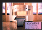
OpenSided MRI
23521 Paseo de Valencia
Suite 113
Laguna Hills, CA 92653
949.587.0093
Pacific Coast
Ultrasound, Inc.
4622 Katella Ave. Suite 101
Los Alamitos, CA 90720
562.596.3428
www.pacificcoastultrasound.com
South County Open MRI
24331 El Toro Rd, Ste 100
Laguna Woods, CA 92653
949.699.6736
Orange
County Diagnostics
27725 Santa Margarita Pkwy, Suite #101
Mission Viejo, CA 92691
949.462.3999
http://ocdiagnostics.com
